Why Spinal Decompression Matters
Spinal decompression therapy is a non‑invasive, computer‑controlled traction technique that gently stretches the spine to create negative pressure within intervertebral discs. By reducing intradiscal pressure, the treatment allows bulging or herniated disc material to retract, restores nutrient flow, and relieves compression of neural elements such as spinal nerves and the spinal cord. Because the procedure is performed on a motorized table with a pelvic harness, it does not require incisions, anesthesia, or medication, making it a drug‑free option that aligns with a patient‑focused, stepped‑care philosophy. The overarching goal is to diminish pain at its source, improve mobility, and enable patients to return to work, school, or daily activities without relying on long‑term opioids or invasive surgery. When combined with complementary therapies—myofascial release, corrective exercises, and ergonomic education—spinal decompression supports lasting functional improvement and a healthier, more resilient spine.
Understanding Spinal Decompression Therapy
![]() Spinal decompression therapy
Spinal decompression therapy is a non‑surgical, motor‑controlled traction treatment that gently stretches the spine to relieve pressure on intervertebral discs and nerve roots. By creating negative pressure within the disc, the technique encourages retraction of bulging material, improves nutrient diffusion, and promotes natural healing. Typical sessions last 30‑45 minutes on a computer‑controlled table and are performed 2‑4 times per week for 4‑6 weeks, usually totaling 12‑28 treatments.
Spinal decompression therapy
Spinal decompression therapy is a non‑surgical, motor‑controlled traction treatment that gently stretches the spine to relieve pressure on intervertebral discs and nerve roots. By creating negative pressure within the disc, the technique encourages retraction of bulging material, improves nutrient diffusion, and promotes natural healing. Typical sessions last 30‑45 minutes on a computer‑controlled table and are performed 2‑4 times per week for 4‑6 weeks, usually totaling 12‑28 treatments.
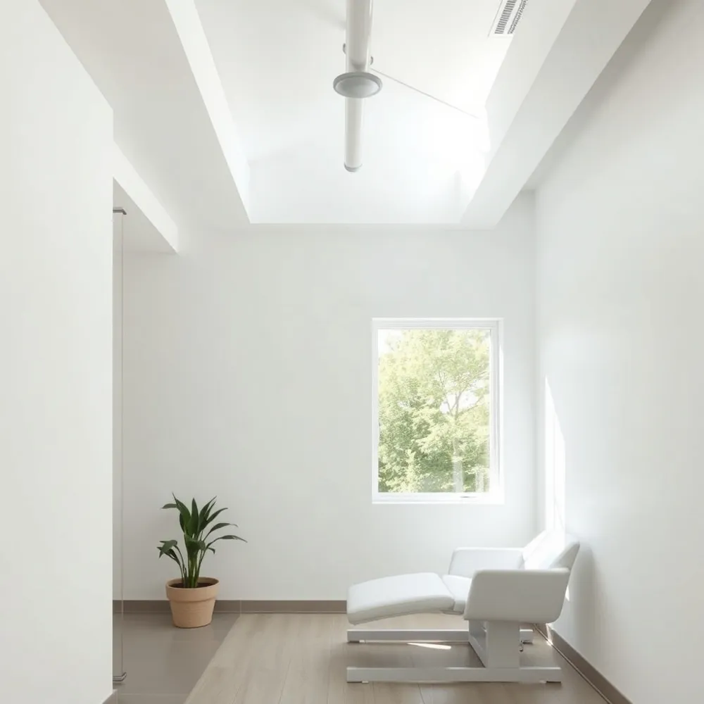
Spinal decompression machine A spinal decompression machine is a motor‑controlled traction device that intermittently pulls on the vertebrae, generating negative intradiscal pressure. The system is computer‑guided, allowing clinicians to customize force, angle, and cycle timing for each patient’s tolerance and therapeutic goal. Popular units include the DRX9000, HillDT, and VAX‑D tables, each equipped with real‑time muscle‑guarding sensors that adjust pull force to prevent guarding and maximize comfort.
Spinal decompression table A spinal decompression table is a motor‑assisted platform with pelvic and trunk harnesses. It delivers precise, rhythmic stretches to the lumbar, cervical, or thoracic spine, creating space between vertebrae that reduces nerve compression. The table’s software programs individualized protocols—often 15‑30 minutes per session—based on imaging findings (MRI, CT, X‑ray) and clinical assessment, ensuring safe and effective treatment for conditions such as herniated discs, degenerative disc disease, sciatica, and spinal stenosis.
Who Is a Good Candidate and Indications
![]() Spinal decompression therapy is most effective for patients whose pain originates from disc‑related compression of nerve roots. Common diagnoses that benefit include herniated or bulging lumbar and cervical discs, degenerative disc disease, lumbar spinal stenosis, and sciatica. These conditions often produce axial or radicular pain, numbness, tingling, and reduced functional mobility that persist after a trial of rest, NSAIDs, heat/cold therapy, physical therapy, or chiropractic adjustments.
Spinal decompression therapy is most effective for patients whose pain originates from disc‑related compression of nerve roots. Common diagnoses that benefit include herniated or bulging lumbar and cervical discs, degenerative disc disease, lumbar spinal stenosis, and sciatica. These conditions often produce axial or radicular pain, numbness, tingling, and reduced functional mobility that persist after a trial of rest, NSAIDs, heat/cold therapy, physical therapy, or chiropractic adjustments.
Imaging and clinical criteria for selection require objective evidence of disc pathology on MRI, CT, or X‑ray, such as a disc bulge, annular tear, or narrowed foramina at the symptomatic level. A thorough clinical exam should reveal corresponding neurologic findings (e.g., positive straight‑leg raise, diminished reflexes) and a history of pain lasting more than four weeks despite conservative care. Patients must be medically stable, able to lie prone for 15‑45 minutes, and willing to commit to a 4‑6‑week protocol of 12‑20 sessions.
Red‑flag conditions that exclude patients are acute spinal fractures, severe osteoporosis, active infection or malignancy, recent spinal surgery (<6 weeks), implanted hardware (pedicle screws/rods), and pregnancy. These contraindications prevent the safe application of traction forces.
Answers to key questions:
- Who is a good candidate for spinal decompression? Good candidates have disc‑related problems such as herniated or bulging discs, degenerative disc disease, or spinal stenosis causing nerve compression, with chronic pain and functional limits after conservative therapy.
- What are the indications for spinal decompression? Indications include persistent nerve‑related symptoms (moderate pain, weakness, numbness) that do not resolve with non‑invasive care, imaging evidence of disc bulge or stenosis, and functional impairment.
- What are the first signs of L4‑L5 compression? Early signs are a dull lower‑back ache, tingling or numbness radiating to the buttocks, hips, or back of the legs, and pain that worsens with standing or walking but improves when sitting or bending forward.
When Not to Use Spinal Decompression
![]() [Spinal decomcompression] is a valuable non‑surgical option for many back‑pain sufferers, but it is not appropriate for everyone. Absolute contraindications include pregnancy, recent spinal fusion or major back surgery (within the past 6 weeks), severe osteoporosis, active infection, tumors, acute vertebral fractures, and significant spinal instability. Patients with implanted hardware (pedicle screws or rods), advanced spondylolisthesis (grade III‑IV), severe scoliosis, or uncontrolled systemic disease also fall into this category.
[Spinal decomcompression] is a valuable non‑surgical option for many back‑pain sufferers, but it is not appropriate for everyone. Absolute contraindications include pregnancy, recent spinal fusion or major back surgery (within the past 6 weeks), severe osteoporosis, active infection, tumors, acute vertebral fractures, and significant spinal instability. Patients with implanted hardware (pedicle screws or rods), advanced spondylolisthesis (grade III‑IV), severe scoliosis, or uncontrolled systemic disease also fall into this category.
Relative contraindications and patient‑specific concerns involve conditions that may increase risk or reduce effectiveness, such as mild‑to‑moderate osteoporosis, chronic anticoagulation, uncontrolled hypertension, or a history of failed back surgery where the anatomy is altered. Each individual’s age, comorbidities, and pain pattern must be weighed against potential benefits.
Importance of professional evaluation: A thorough history, physical exam, and imaging (MRI, CT, or X‑ray) performed by a qualified chiropractor, physiatrist, or spine specialist is essential before initiating therapy. This assessment identifies red‑flag signs, confirms that the disc pathology is amenable to traction, and ensures safety.
When should you not do spinal decompression? The therapy should be avoided in anyone who is pregnant, has recent fusion or major surgery, severe osteoporosis, infection, tumor, fracture, or marked instability; also in patients with certain hardware or severe deformities.
Is it safe to do minimally invasive lumbar decompression on a 90‑year‑old? Age alone does not preclude the procedure, but elderly patients have higher risk for complications. A comprehensive health assessment, imaging, and discussion of comorbidities are required to determine suitability.
Does spinal decompression make you taller? The treatment can temporarily increase height by a quarter to half an inch as discs re‑hydrate and separate, but the effect is short‑lived; the true benefit lies in reduced pain, improved posture, and enhanced mobility.
Benefits, Risks, and Success Rates
![]() Spinal decompression therapy offers a non‑invasive, drug‑free option that gently stretches the spine to relieve pressure on discs and nerve roots. The major advantages are that it can reduce nerve irritation, improve mobility, and allow patients to avoid surgery while staying fully clothed during treatment. It is customizable to each patient’s tolerance, can be combined with adjunctive modalities such as myofascial release, corrective exercises, and custom orthotics, and is generally safe for healthy adults. However, the therapy is not appropriate for people with severe osteoporosis, acute fractures, spinal infections, tumors, implanted hardware, or pregnancy, and it requires a commitment to multiple 15‑45‑minute sessions over four to six weeks. Insurance coverage varies, and long‑term scientific evidence is still limited. Clinical reports from a variety of clinics and research studies cite success rates ranging from 70 % to 86 % for pain relief and functional improvement, with some large case series documenting up to 88 % of patients experiencing significant benefit after a full course. Long‑term durability is mixed; many patients maintain relief for months to years when they continue posture correction, core‑strengthening, and periodic maintenance sessions, but the therapy is not a permanent cure and recurrence can occur, especially if underlying degenerative changes progress.
Spinal decompression therapy offers a non‑invasive, drug‑free option that gently stretches the spine to relieve pressure on discs and nerve roots. The major advantages are that it can reduce nerve irritation, improve mobility, and allow patients to avoid surgery while staying fully clothed during treatment. It is customizable to each patient’s tolerance, can be combined with adjunctive modalities such as myofascial release, corrective exercises, and custom orthotics, and is generally safe for healthy adults. However, the therapy is not appropriate for people with severe osteoporosis, acute fractures, spinal infections, tumors, implanted hardware, or pregnancy, and it requires a commitment to multiple 15‑45‑minute sessions over four to six weeks. Insurance coverage varies, and long‑term scientific evidence is still limited. Clinical reports from a variety of clinics and research studies cite success rates ranging from 70 % to 86 % for pain relief and functional improvement, with some large case series documenting up to 88 % of patients experiencing significant benefit after a full course. Long‑term durability is mixed; many patients maintain relief for months to years when they continue posture correction, core‑strengthening, and periodic maintenance sessions, but the therapy is not a permanent cure and recurrence can occur, especially if underlying degenerative changes progress.
Surgical Decompression vs. Non‑Surgical Options
![]() When surgery becomes necessary
Patients who have chronic radicular pain, progressive neurological deficits, or disabling spinal stenosis that does not improve after 4‑6 weeks of conservative care (physical therapy, NSAIDs, chiropractic adjustments, traction, or TENS) are considered for surgical decompression. Imaging (MRI, CT, X‑ray) must confirm disc herniation, severe stenosis, or bone spur that compresses nerve roots.
When surgery becomes necessary
Patients who have chronic radicular pain, progressive neurological deficits, or disabling spinal stenosis that does not improve after 4‑6 weeks of conservative care (physical therapy, NSAIDs, chiropractic adjustments, traction, or TENS) are considered for surgical decompression. Imaging (MRI, CT, X‑ray) must confirm disc herniation, severe stenosis, or bone spur that compresses nerve roots.
Common surgical procedures and their goals
- Laminectomy: removes the lamina to enlarge the spinal canal and relieve pressure on the cord or nerve roots.
- Laminotomy: a partial lamina removal targeting a specific nerve exit.
- Foraminotomy: enlarges the foramen to free a pinched nerve.
- Laminoplasty: reshapes the lamina while preserving stability.
- Micro‑discectomy: excises the protruding disc fragment causing radiculopathy. These techniques aim to restore space, reduce inflammation, and improve functional mobility.
Relative seriousness and recovery considerations Surgical decompression is invasive, carries risks (infection, bleeding, blood clots, dural tears, nerve injury, spinal instability), and typically requires weeks to months of recovery with activity restrictions and physical therapy. The decision balances potential long‑term relief against these risks.
How serious is spinal decompression surgery? Spinal decompression surgery is an invasive procedure reserved for severe or persistent nerve compression when conservative care fails. Risks include infection, bleeding, blood clots, dural tears, nerve injury, and spinal instability. Recovery may take weeks to months, and the decision is based on a thorough evaluation of benefits versus risks.
Types of spinal decompression surgery Common types are laminectomy (removing the lamina), laminotomy (partial lamina removal), foraminotomy (enlarging the nerve exit), laminoplasty (hinged lamina), micro‑discectomy, and endoscopic decompression. Choice depends on location, cause of compression, and patient health.
Is chiropractic spinal decompression effective? Yes. Chiropractic spinal decompression, when performed by a qualified chiropractor, effectively reduces nerve pressure and alleviates pain from herniated or bulging discs, sciatica, and related conditions. It is safe for most individuals, though contraindicated for pregnancy, tumors, metal implants, severe osteoporosis, fractures, and certain vascular issues.
Practical Considerations: Cost, Insurance, Sessions, and Duration
![]() How much should spinal decompression cost?
How much should spinal decompression cost?
In the United States a typical session ranges from $50 to $150. Because most patients require a series of treatments—often 10‑20 sessions—clinics frequently offer package discounts or financing plans. After an initial chiropractic evaluation, Dr. Allison Ross (San Jose) provides a personalized estimate based on the prescribed protocol.
Is spinal decompression covered by insurance?
Coverage varies. Many insurers label mechanical spinal decompression as experimental, so it is not automatically reimbursed. However, if the therapy is documented as medically necessary and performed by an in‑network provider, some plans will pay partially. Medicare generally does not cover standalone decompression, although manual traction may be covered under certain conditions.
How many sessions do you need for spinal decompression?
A standard program at Ross Chiropractic includes 8‑12 sessions for noticeable relief, with many patients reporting improvement after 4‑6 visits. More chronic or severe cases may require 15‑20 sessions spread over four to six weeks.
How long does spinal decompression last?
Therapy is typically delivered over a four‑to‑six‑week period. Pain reduction often begins within the first few treatments and can persist for weeks to months after the course. Maintaining proper posture, core‑strengthening exercises, and occasional maintenance sessions can extend benefits to three‑to‑six months or longer.
Home Care, Exercises, and Lifestyle Adjustments
![]() After a clinic‑based spinal decompression program, patients can extend the benefits at home with gentle, low‑impact stretches that mimic the traction forces used on the table. Simple spinal elongation moves such as Child’s Pose, Knees‑to‑Chest, Cat‑Cow, Prone Pillow Decompression Stretch, and back‑extension rolls on an exercise ball provide a mild negative pressure that encourages disc re‑hydration and reduces nerve irritation. Core‑strengthening exercises like dead‑bug, bird‑dog, and modified planks support the lumbar spine and help maintain the space created during therapy.
After a clinic‑based spinal decompression program, patients can extend the benefits at home with gentle, low‑impact stretches that mimic the traction forces used on the table. Simple spinal elongation moves such as Child’s Pose, Knees‑to‑Chest, Cat‑Cow, Prone Pillow Decompression Stretch, and back‑extension rolls on an exercise ball provide a mild negative pressure that encourages disc re‑hydration and reduces nerve irritation. Core‑strengthening exercises like dead‑bug, bird‑dog, and modified planks support the lumbar spine and help maintain the space created during therapy.
For those focused on L4‑L5 relief, a side‑lying knee‑to‑chest stretch, an overhead side‑bend, or a brief inversion (30‑60 seconds) on a pull‑up bar can safely decompress the lower lumbar segments. Perform each movement slowly, stop at any pain, and verify the routine with a qualified practitioner such as Dr. Ross.
Avoid activities that aggravate L5 compression: heavy deadlifts, deep squats, high‑impact running or jumping, and repetitive twisting motions. These increase intradiscal pressure and can reverse therapeutic gains.
A common myth is that spinal decompression permanently adds height. In reality, re‑hydrated discs may temporarily add ½‑1 inch, but the true advantage is improved posture, reduced pain, and better functional mobility, not a lasting increase in stature.
Safety Measures, Patient Education, and Long‑Term Wellness
![]() Clinics that offer spinal decompression therapy must follow strict safety protocols and use FDA‑cleared equipment such as the DRX9000 or HillDT tables. Devices are inspected regularly, and emergency stop switches are installed for both clinician and patient use. Before a session begins, a qualified chiropractor or physical therapist conducts a comprehensive evaluation, reviewing medical history, imaging (MRI, CT or X‑ray) and confirming that no contraindications—such as acute fractures, severe osteoporosis, infection, malignancy, spinal implants, or pregnancy are present.
Clinics that offer spinal decompression therapy must follow strict safety protocols and use FDA‑cleared equipment such as the DRX9000 or HillDT tables. Devices are inspected regularly, and emergency stop switches are installed for both clinician and patient use. Before a session begins, a qualified chiropractor or physical therapist conducts a comprehensive evaluation, reviewing medical history, imaging (MRI, CT or X‑ray) and confirming that no contraindications—such as acute fractures, severe osteoporosis, infection, malignancy, spinal implants, or pregnancy are present.
During treatment, patients wear comfortable, non‑restrictive clothing and are securely harnessed to the motorized table. Proper positioning and a snug pelvic harness ensure even traction while minimizing the risk of muscle guarding. Clinicians maintain open communication, asking patients to report any new or worsening discomfort promptly so that traction forces can be adjusted in real time.
After each session, patients receive clear post‑treatment guidance: stay well‑hydrated, avoid heavy lifting or twisting for several hours, and follow a prescribed home‑exercise program that includes core strengthening, posture correction, and gentle stretches. Follow‑up appointments are scheduled to reassess pain scores, functional mobility and imaging results, allowing the care plan to be refined and long‑term spinal health to be maintained.
Putting It All Together
Spinal decompression therapy works best when it is delivered as a personalized, step‑wise program that respects each patient’s unique anatomy and symptom pattern. After a thorough chiropractic or medical evaluation—including imaging such as MRI or CT—clinicians rank treatment options from least to most invasive, beginning with rest, heat, NSAIDs, and targeted exercises, then progressing to gentle traction, myofascial release, and finally the motor‑controlled decompression table if pain persists. Throughout this ladder, clinicians constantly weigh the therapy’s proven benefits—pain relief, disc re‑hydration, faster return to work—against absolute contraindications such as acute fracture, severe osteoporosis, infection, malignancy, or pregnancy. By integrating adjunctive strategies (core‑strengthening, ergonomic education, custom orthotics) and encouraging patients to adopt healthy posture and activity habits, the program promotes lasting spinal health far beyond the treatment window, reducing recurrence and the need for future surgery. Patients often report improved quality of life and sustained functional gains.